Cell and Tissue Imaging Core
The overall objective of the Cell and Tissue Imaging Core is to provide an integrated approach to investigate the structure and dynamic behavior of diabetes-related cells and tissues. By providing access to and technical support in using advanced cellular microscopy tools, the Cell and Tissue Imaging Core will accelerate the pace, expand the scope, and improve efficiency of diabetes research.
Advanced cellular microscopy is a powerful tool for biological research and has an important role to play in the study of the pathogenesis and treatment of diabetes mellitus. Imaging technology has evolved rapidly over the last decade leading to improvements in resolution, sensitivity and speed which have created fundamentally new opportunities for studying processes across many orders of magnitude and in real-time in living cells and animals. At the same time, the costs of increasingly sophisticated equipment are substantial and the expertise to efficiently use, maintain, and develop this equipment is not common in most labs. The overall objective of the Cell and Tissue Imaging Core is to provide an integrated approach to investigate the structure and dynamic behavior of diabetes-related cells and tissues. By providing access to and technical support in using advanced cellular microscopy tools, the Cell and Tissue Imaging Core will accelerate the pace, expand the scope, and improve efficiency of diabetes research.
Leadership

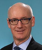
David Piston, PhD, Interim Director
E Mallinckrodt Jr Professor & Chair of Cell Biology & Physiology
660 South Euclid Avenue, MSC 8228-0003-04
St. Louis, MO 63110
Phone 314-362-9121
Email piston@wustl.edu
Other Contacts
Dennis Oakley, BS,
Research Specialist, light microscopy
660 South Euclid Avenue, MSC 8108-0041-0B
St Louis, MO 63110
Phone 314-747-0870
Email oakleyd@wustl.edu
Michael Chien-cheng Shih, PhD, Research Specialist, light microscopy
660 South Euclid Avenue, Campus Box 8108
St Louis, MO 63110
Phone 314-362-3446
Email c.shih@wustl.edu
Matthew Joens, BS, Research Specialist, electron microscopy
660 South Euclid Avenue, Campus Box 8108
St Louis, MO 63110
Phone 314-362-0838
Email mjoens@wustl.edu
Robyn Roth, BS, Research Specialist, electron micrsocopy
660 South Euclid Avenue, MSC 8228-0002-04
St. Louis, MO 63110
Phone 314-747-1610
Email rroth22@wustl.edu
Karen Green, BA, Research Specialist, electron microscopy
660 South Euclid Avenue, MSC 8118-0014-04
St. Louis, MO 63110
Phone 314-362-7462
Email greenkg@wustl.edu
Services
Light Microscopy
- Routine Microscopy: Both upright and inverted microscope platforms capable of both Differential Interference Contrast (DIC) and multi-color fluorescence (DAPI/FITC/TRITC/Cy5) of fixed samples.
- Slide Scanning Microscopy: An automated slide-scanning platform capable of scanning complete slides in either bright-field or multi-color fluorescence (DAPI/FITC/TRITC/Cy5) modes. System is equipped with a slide autoloader and automation software.
- Confocal Microscopy: An upright confocal microscope platform capable of both transmitted light Differential Interference Contrast (DIC) and multi-color fluorescence imaging of fixed mounted cell cultures or tissue sections as well as living specimens such as C. elegans and zebrafish model systems. The system is also equipped with a second scanner for photo-bleaching experiments and photo-stimulation paradigms such as optogenetics.
- Live-cell Microscopy: Two inverted confocal microscope platforms for 3D imaging of living specimens equipped with live-cell incubation chambers capable of controlling CO2, O2, humidity and temperature. The first is a resonant scanning LSM equipped with six laser lines (405, 445, 480, 515, 561 and 640nm) capable of spectral and FRET imaging, the second is a spinning disk confocal equipped with four laser lines (405, 480, 561 and 640nm) and two sCMOS cameras capable of simultaneous two-color imaging.
- TIRF / STORM Microscopy: An inverted platform equipped with live-cell incubation chambers capable of controlling CO2, O2, humidity and temperature and a motorized TIRF illuminator with four laser times (405, 480, 561 and 640nm) and two cameras. One camera (sCMOS) is designated for high-speed TIRF imaging, the other camera (EM-CCD) is designated for single-molecule imaging, specifically STOchastic Reconstruction Microscopy that enables imaging beyond the diffraction limit of light at ~20-40nm resolution in XY and 100nm in Z.
- SIM Super-Resolution Microscopy: An inverted platform equipped with four laser times (405, 480, 561 and 640nm) and two EM-CCD cameras capable of simultaneous two-color imaging at twice the resolution of confocal microscopy of fixed mounted cell cultures and thin tissue sections.
- Two-Photon Microscopy: An inverted two-photon microscope capable of multi-color fluorescence imaging of fixed mounted cell cultures or tissue sections as well as live specimens such as in vivo mouse imaging. The system is equipped with a Coherent Discovery laser for two-photon excitation and Gallium Arsenide Phosphide (GaAsP) detectors for high-sensitivity detection of fluorescence signals.
- Training and Consultation, Image acquisition: Consultation includes experimental planning, technical advice on cell and tissue fixation, and discussion of appropriate fluorescent reporter probes. Training is performed either by Mr. Oakley or Dr. Shih. The users are trained to examine their own tissue, capture images and discuss findings with Dr. Fitzpatrick and the WUCCI LM research specialists.
Electron Microscopy (EM)
- Routine tissue EM: Service includes processing and embedding (maximum of 6 blocks), cutting 1 micron thick plastic sections stained with toluidine blue, cutting of thin sections on standard 200 mesh grids, post-staining in uranyl acetate and lead citrate, and consultation.
- Tissue culture EM: Service includes routine procedures described above but requires additional specimen preparation and cutting time.
- Immuno-EM: Services include LR White embedding media or pre-embed immunostaining, processing and embedding (maximum of 6 blocks) or cutting 1 micron plastic sections on slides – stained for toluidine blue. Additional services include cutting thin sections on grids, primary antibody staining (primary antibody supplied by investigator), secondary antibody gold labeling (secondary antibody charged to investigator), counterstaining with OsO4, uranyl acetate, lead citrate, and consultation.
- Deep-Etch-EM: Service includes slam freezing of samples in liquid helium, freeze etching and free fracture as well as platinum replica production onto TEM grids.
- Negative Staining: Services include preparation of formvar-coated grids and negative staining techniques appropriate for the virus or particle to be visualized.
- Metal coating: Service includes critical point drying of samples and subsequent metal coating for topographic imaging and 3DEM in the SEM and FIB-SEM microscope platforms.
- Training and Consultation, Image acquisition: Consultation includes experimental planning, technical advice on tissue fixation, and discussion of appropriate ultrastructural analysis. Ms. Roth performs TEM imaging and SEM, and FIB-SEM 3DEM imaging is performed by Mr. Joens. Both Ms. Roth and Mr. Joens examine the respective tissue samples with the investigator/trainee. Discussion of findings is undertaken with Dr. Fitzpatrick and the WUCCI EM research specialists as required.
X-ray Microscopy
- X-ray Microscopy: A Zeiss Xradia 520 Versa X-ray microscope is available on a collaborative basis for the sub-micron tomographic imaging of tissues samples from microns to many centimeters in size.
- Training and Consultation, Image acquisition: Consultation includes experimental planning, technical advice on cell and tissue fixation, and discussion of appropriate contrast agents. Dr. Fitzpatrick performs the imaging as well as leads any subsequent discussion of results.
Getting Started
Consultation with Dr. Piston and WUCCI staff is available and encouraged for all PIs and their laboratory members to plan experiments, select imaging modalities, and interpret data.
iLab on-line scheduling for trained users
Chargebacks and DRC Scholarships
User fees are charged for microscope time and assistance in order to recover partial costs of service contracts, supplies including computers, software, minor upgrades and general maintenance (Table 1). Such fees are based on the actual direct costs required to perform the service(s). The WUCCI IAC reviews costs, usage and expenditures twice annually and user fees are adjusted as necessary in accordance with Washington University and NIH guidelines for operation of a large recharge center. Should any excess funds be generated, they will be used to lower fees charged to users.
Fee Schedule for Light Microscopy Services
| Service | Peak (Mon-Fri 9am-5pm) | Off-peak (nights, weekends) |
| Fluorescence/Bright-Field Microscopy | $20.00 per hour | $10.00 per hour |
| Slide Scanning Microscopy | $25.00 per hour | $15.00 per hour |
| Confocal Microscopy | $30.00 per hour | $20.00 per hour |
| Live-cell Microscopy | $30.00 per hour | $20.00 per hour |
| Long-term daily charge (24 hr) | $300.00 per day | |
| TIRF/STORM Microscopy | $30.00 per hour | $20.00 per hour |
| SIM Microscopy | $30.00 per hour | $20.00 per hour |
| Two-Photon Microscopy | $30.00 per hour | $20.00 per hour |
| Initial Training | $150 one time cost | |
| Miscellaneous Charges | ||
| Technical Specialist Time * | $60.00 per hour with a half hour minimum |
* Technical fees are built into cost of each service. Additional technical time is charged for services not listed above, including “imaging for hire” when WUCCI staff acquire data.
Fee Schedule for Electron Microscopy Services
| Service | Peak (Mon-Fri 9am-5pm) |
| Deep-Etch EM | $250 per sample |
| Embedding / staining / sectioning | $180 per sample |
| High-pressure freezing | $230 per sample |
| Immuno-labeling run (up to 6 samples) | $250 one time cost |
| Routine sectioning | $90.00 per hour |
| Cryo-sectioning | $120.00 per hour |
| FIB-SEM sample preparation | $180.00 per sample |
| Metal (Au, Pd, Pt, Ir) / Carbon Coating | $30.00 per run |
| Negative Stain | $15.00 per grid |
| TEM time (assisted) | $75.00 per hour |
| SEM / FIB-SEM (assisted) | $65.00 per hour |
| Long-term daily charge (24 hr) | $500.00 per day |
| Miscellaneous Charges | |
| Technical Specialist Time * | $60.00 per hour with a half hour minimum |
* Technical fees are built into cost of each service. Additional technical time is charged for services not listed above, including “imaging for hire” when WUCCI staff acquire data.
Fee Schedule for X-Ray Microscopy Services
| Service | Peak (Mon-Fri 9am-5pm) | Off-peak (nights, weekends) |
| Xradia Versa 520 XRM | $60.00 per hour | $40.00 per hour |
| Long-term daily charge (24 hr) | $600 per day | |
| Data Analysis | ||
| High-end Data Analysis Workstation | $10.00 per hour | $5.00 per hour |
| Long-term daily charge (24 hr) | $100 per day | |
| Miscellaneous charges | ||
| Technical Specialist Time * | $60.00 per hour with a half hour minimum |
* Technical fees are built into cost of each service. Additional technical time is charged for services not listed above, including “imaging for hire” when WUCCI staff acquire data.
DRC funding of the Cell and Tissue Imaging Core contributes to lower costs for use by DRC members. By university and NIH policy, all users are subject to the same chargebacks. However, a portion of those chargebacks are credited back to DRC investigators via scholarships, effectively decreasing the financial burden to DRC members. Scholarships of $500 (for smaller projects) or $1000 (for larger projects) will be granted on a first-come, first-served basis, with the goal of scholarship support covering 50% of the expected chargebacks for expected project expenses.
Scholarship funding is currently available for FY23. Please contact Interim Core Director, Dr. Piston for more information at piston@wustl.edu.
You must be logged in to post a comment.